Back Muscles Chart : Anatomy For Athletes Part 7 Intrinsic Back Muscles Formidable Female Fitness. Superficial, intermediate, deep and deepest layers.these muscles lie on each side of the vertebral column, deep to the thoracolumbar fascia they span the entire length of the vertebral column, extending from the cranium to the pelvis The teres major is a small, yet important muscle within the back. Backbone diagram with vertebrae, disks and nerves. To download your free copy click the link. Anatomynote.com found anatomy of back muscles diagram from plenty of anatomical pictures on the internet.
The superficial group, the deep group, and the intermediate group. To download your free copy click the link. Claim your free copy of the client back care guide today. Certain back muscles extend to other areas, like the shoulders, upper arms, and thighs. If you experience any of these symptoms, seek medical attention immediately.
Chart Human Anatomy And Physiology Of Muscles from www.anatomy.link Adductor magnus biceps femoris carpi flexor ulnaris deltoid. We think this is the most useful anatomy picture that you need. Raises and rotates arm in all directions. Sciatica medical health care vector illustration scheme with lower spine and sciatic nerve pain in leg. Your clients will thank you for it! If you experience any of these symptoms, seek medical attention immediately. We hope this picture anatomy of back muscles diagram can help you study and research. The muscles on each side form a trapezoid shape.
These muscles include the large paired muscles in the lower back, called erector spinae, which help hold up the spine, and gluteal muscles.
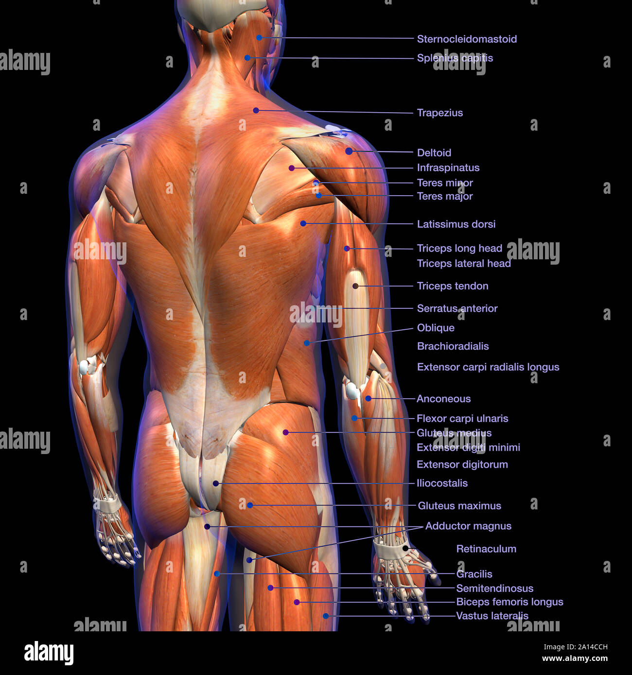
Symptoms of muscle pain include: The muscles on each side form a trapezoid shape. The trapezius and latissimus dorsi muscles connect the upper limb to the vertebral column. We've created a free trigger point chart, which includes fybromyalgia treatment and reflexology information. The teres major is a small, yet important muscle within the back. The fibres attach to the clavicle, acromion and the scapula spine. Your clients will thank you for it! Musculoskeletal image with rounded rectangle on white background. Muscle charts of the human body for your reference value these charts show the major superficial and deep muscles of the human body. Muscle anatomy chart 12 photos of the muscle anatomy chart abdominal muscle anatomy chart, human muscle anatomy diagram free, interactive muscle anatomy chart, pelvic muscle anatomy chart, shoulder muscle anatomy chart, human muscles, abdominal muscle anatomy chart, human muscle anatomy diagram free. There are three different muscle groups found in the back: Artery) p.134 accessory nerve p. Backbone diagram with vertebrae, disks and nerves.
Anatomynote.com found anatomy of back muscles diagram from plenty of anatomical pictures on the internet. The muscles of the lower back help stabilize, rotate, flex, and extend the spinal column, which is a bony tower of 24 vertebrae that gives the body structure and houses the spinal cord.the spinal. We hope this picture anatomy of back muscles diagram can help you study and research. It is attached to the calcaneus and is pulled by 3 flexor muscles: Certain back muscles extend to other areas, like the shoulders, upper arms, and thighs.
Intrinsic Back Muscles Anatomy Of The Torso Medical Library from d3uigcfkiiww0g.cloudfront.net The teres major is a small, yet important muscle within the back. An extremely strong tendon attached to the heel. The fibres attach to the clavicle, acromion and the scapula spine. Extends spine and trunk back. The superficial group, the deep group, and the intermediate group. The soleus, the plantaris, and the gastrocnemius. Certain back muscles extend to other areas, like the shoulders, upper arms, and thighs. The vast majority of back problems improve on their own or with nonsurgical treatment.
The soleus, the plantaris, and the gastrocnemius.
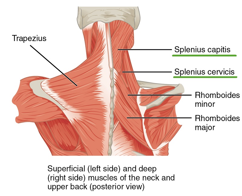
Muscle anatomy chart 12 photos of the muscle anatomy chart abdominal muscle anatomy chart, human muscle anatomy diagram free, interactive muscle anatomy chart, pelvic muscle anatomy chart, shoulder muscle anatomy chart, human muscles, abdominal muscle anatomy chart, human muscle anatomy diagram free. Try a few basic exercises to stretch and strengthen your back and supporting muscles. Muscles found in the superficial group include rhomboid major, rhomboid minor, levator scapulae, trapezius, latissimus dorsi. Three types of back muscles that help the spine function are extensors, flexors and obliques. The extrinsic (superficial) back muscles, which lie most superficially on the back. Muscle action 12 photos of the back muscle chart. 1) make midline incision along spines of vertebrae 2) extend from Back anatomy stock photos and images. A strain can be an injury to a tendon attachment from muscle to bone. Your clients will thank you for it! An extremely strong tendon attached to the heel. For more anatomy content please follow us and visit our website: Muscle charts of the human body for your reference value these charts show the major superficial and deep muscles of the human body.
The superficial group, the deep group, and the intermediate group. Three types of back muscles that help the spine function are extensors, flexors and obliques. Muscle charts of the human body for your reference value these charts show the major superficial and deep muscles of the human body. Back anatomy stock photos and images. Other muscles are small and cover much less space.
Labeled Anatomy Chart Of Male Back Muscles On Black Background Stock Photo Alamy from c8.alamy.com Anatomy chart courtesy of fcit the latissimus dorsi muscles (also known as the lats) are the largest muscles of the back. Strain commonly occurs with incorrect lifting of heavy. The rhomboid muscle is activated as you bring and squeeze your scapula or shoulder blades back and together. Keep your torso upright and a slight arch in your back as you fully extend your arms at the top. The trapezius and latissimus dorsi muscles connect the upper limb to the vertebral column. Back anatomy stock photos and images. The vast majority of back problems improve on their own or with nonsurgical treatment. Leaning back to straight vertical and all points in between.
For images of the muscle, click on each link under location.
Keep your torso upright and a slight arch in your back as you fully extend your arms at the top. For more anatomy content please follow us and visit our website: Adductor magnus biceps femoris carpi flexor ulnaris deltoid. This procedure is one of the most powerful yet simple ways to treat muscle pain and discomfort. The teres major is a small, yet important muscle within the back. Back anatomy stock photos and images. If you experience any of these symptoms, seek medical attention immediately. It is the most superficial of all the back muscles. Some of these muscles are quite large and cover broad areas. The muscles of the back are a group of strong, paired muscles that lie on the posterior aspect of the trunk they provide movements of the spine, stability to the trunk, as well as the coordination between the movements of the limbs and the back muscles are divided into two large groups: The trapezius and latissimus dorsi muscles connect the upper limb to the vertebral column. Overview product description the muscles of the shoulder and back chart shows how the many layers of muscle in the. Certain back muscles extend to other areas, like the shoulders, upper arms, and thighs.